ctDNA, CTCs and the Future of Liquid Biopsy
Overcoming challenges with novel technology and a tale of two halves?
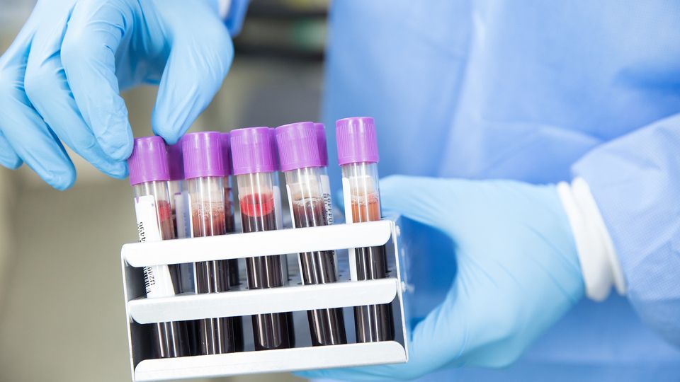
Liquid biopsy (LBx) has emerged as a transformative tool in oncology, offering a minimally invasive means to access real-time molecular insights into tumor biology. By analyzing components shed from tumors into the bloodstream – particularly circulating tumor DNA (ctDNA) and circulating tumor cells (CTCs) – LBx provides unprecedented opportunities for early detection, treatment stratification, therapeutic monitoring and the identification of resistance mechanisms. While ctDNA offers a dynamic snapshot of the tumor’s genomic landscape, CTCs represent intact cellular material that can yield critical phenotypic and functional data, including RNA transcription, protein markers and cellular behavior.
As the clinical and research communities have deepened their reliance on LBx, the integration of ctDNA and CTC analyses is becoming increasingly important. However, technological limitations, particularly around the isolation and characterization of rare CTC populations, have historically posed significant barriers. Recent advancements in microfluidics, single-cell analysis and next-generation sequencing (NGS) are beginning to overcome these challenges, opening new frontiers in cancer diagnostics and personalized medicine.
We spoke to Dr. Adam Corner, market development manager, Dr. Yoon-Tae Kang, staff scientist – market development and applications, and Dr. Aqila Ahmed, sales specialist – market development from BioRad, at the American Association of Cancer Research (AACR) Annual Meeting 2025. They shared insights on the challenges of CTCs and ctDNA as cancer diagnostic tools, how novel technology is helping to overcome some of these issues and the future monitoring landscape.
What are the biggest challenges in cancer diagnosis and ongoing monitoring using CTCs and ctDNA?
Adam Corner, PhD (AC):
For diagnosis and monitoring in cancer, the biggest issues are sensitivity and availability of LBx material. Although LBx is described as a minimally invasive sampling method and is relatively easy to obtain compared to tumor biopsies, volumes are naturally limited and obtaining sufficient quantities for deep monitoring of early cancer and residual disease is challenging. This can be overcome with technologies that allow monitoring of multiple markers per genome, e.g., broad whole genome sequencing or whole exome sequencing for tens to hundreds of single nucleotide variants. Compared to NGS technologies, however, being able to monitor many markers in a multiplex droplet digital polymerase chain reaction (ddPCR) can overcome the limited sensitivity of NGS to some extent and combines naturally with a simpler workflow and shorter turnaround time.
Yoon-Tae Kang, PhD (YTK):
CTCs have similar challenges around sample volume, given their limited frequency per blood volume. Broad antibody profiling can still be achieved with newer antibody types and broader spectral microscope capabilities. Accessing the genomic, proteomic as well as metabolomic contents of CTCs is a growing area of interest and may allow for the combined ctDNA and CTC (multiomics) approach to further enhance the power of LBx.
KS:
Aqila Ahmed, PhD (AA):
CTC broadly describes cells that have sloughed off a (typically) primary tumor and are circulating within the blood system. Identifying them can be challenging due to their potential for dynamic phenotypes and well-known epithelial-to-mesenchymal transition (EMT) during metastasis.
Classically, identifying CTCs in a background of white blood cells (WBCs) involves immunofluorescent staining using an anti-CD45 antibody to negatively select WBCs, and an anti-pancytokeratin antibody to positively stain CTCs. This can potentially misidentify other circulating markers such as extracellular vesicles and is dependent on the specificity and sensitivity of the antibody reagents. Therefore, captured cell types are often further stained with established surface markers such as EpCAM, vimentin or PD-L1.
However, not all CTCs express the same surface marker profile, nor do they maintain a constant profile during their individual cellular journey. For example, head and neck squamous cell carcinomas do not typically express EpCAM, and breast cancer CTCs may lose EpCAM and gain vimentin as they progress through the EMT process. So, the use of surface markers to select CTCs initially is fraught with decisions that may subsample the overall patient CTC population, adding complexity that needs further research to have maximum benefit to patients.
YTK:
The Genesis system with Celselect slides format offers label-free cell isolation using differences in physical size and deformity between target cells and non-target cells, such as WBCs, to allow enrichment of CTCs from whole blood. This approach will select potential cancer cells regardless of their antigen presentations within a liquid biopsy sample and enable slide staining or enrichment for various downstream analysis processes, including single-cell RNA sequencing and/or multiplex ddPCR assays for mutation analysis, molecular residual disease (MRD) and drug efficacy. The Genesis system is also compatible with new downstream approaches in the field such as single cell protein sequencing and metabolite studies of captured CTCs thanks to its high sensitivity and high cell viability after processing.
KS:
YTK:
The size-based selection capabilities of the Genesis system with Celselect Slides technology are amenable to capturing and enriching any cell within the LBx sample above a certain size limit. The benefits of this approach are that it does not require prior knowledge of the cell surface marker profile of suspected cancer cells, is a gentle method that retains cell viability and is suitable for CTC studies in less-researched cancer types.
KS:
AC:
As technologies advance, it enables parallel analysis of both ctDNA and CTCs from the same LBx sample, providing comparisons in multiple facets. Smilkou et al (2025) provide one of the first illustrations of a potential lack of correlation between ctDNA and CTC gDNA. More follow-up research is required to demonstrate the value of potentially monitoring multiple analytes from the same LBx sample. It is also possible that each marker tells a different story and reflects a different part of ongoing cancer status. It is exciting work with the potential to impact how LBx are collected and utilized in a clinical setting, but so far this has only been done in a research/translational environment. Being able to identify resistance mutations in CTCs that are not detectable in comparable ctDNA could potentially impact therapeutic decisions.
KS:
Where do you see future developments in monitoring moving towards?
AC:
Current molecular monitoring paradigms are primarily focused on tumor-informed, personalized profiles of mutations via NGS or ddPCR. This can provide a sensitive and specific monitoring capability for individual patients post curative intent treatments i.e., MRD. However, upfront NGS of individual patient samples is costly and time consuming.
With an expansion in epigenetic profiling of cancer genomes and the potential identification of cancer type-specific methylation profiles, the detection and monitoring of early cancer and residual disease could become less personalized and more turnkey. There is potential for the development of methylation profiles per cancer type that can then be used to monitor all patients with a particular type of cancer. In parallel, the development of more user-friendly methylation-based approaches beyond bisulphite conversion, for example, methylation-sensitive restriction enzyme-ddPCR will make this amenable to standard laboratory testing.


